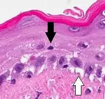
Vacuolar interface dermatitis, with lymphocytes in the dermis and epidermis (black arrow indicates one), and vacuolization (white arrow) at the dermoepidermal junction.
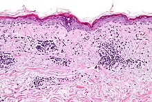
Micrograph of a vacuolar interface dermatitis with dermal mucin, as may be seen in lupus. H&E stain.
Vacuolar interface dermatitis (VAC, also known as liquefaction degeneration, vacuolar alteration or hydropic degeneration) is a dermatitis with vacuolization at the dermoepidermal junction, with lymphocytic inflammation at the epidermis and dermis.[1]
Causes
| Main conditions[2] | Characteristics | Micrograph | Photograph | |
|---|---|---|---|---|
| Generally/Not otherwise specified | Typical findings, called "vacuolar interface dermatitis":[2]
|
|
||
| Acute graft-versus-host-disease |  |
|||
| Allergic drug reaction |  |
|||
| Lichen sclerosus | Hyperkeratosis, atrophic epidermis, sclerosis of dermis and dermal lymphocytes.[3] |  | ||
| Erythema multiforme | ||||
| Lupus erythematosis | Typical findings in systemic lupus erythematosus:[4]
|
 |
 |
An interface dermatitis with vacuolar alteration, not otherwise specified, may be caused by viral exanthems, phototoxic dermatitis, acute radiation dermatitis, erythema dyschromicum perstans, lupus erythematosus and dermatomyositis.[2]
References
- ↑ Bolognia, Jean L.; et al. (2007). Dermatology. St. Louis: Mosby. p. 11. ISBN 1-4160-2999-0.
- 1 2 3 4 5 6 7 Unless else specified in boxes, reference is: Alsaad, K O (2005). "My approach to superficial inflammatory dermatoses". Journal of Clinical Pathology. 58 (12): 1233–1241. doi:10.1136/jcp.2005.027151. ISSN 0021-9746.
- ↑ Lisa K Pappas-Taffer. "Lichen Sclerosus". Medscape. Updated: May 17, 2018
- ↑ Mowafak Hamodat. "Skin inflammatory (nontumor) > Lichenoid and interface reaction patterns > Lupus: systemic lupus erythematosus (SLE)". PathologyOutlines. Topic Completed: 1 August 2011. Revised: 26 March 2019
This article is issued from Wikipedia. The text is licensed under Creative Commons - Attribution - Sharealike. Additional terms may apply for the media files.